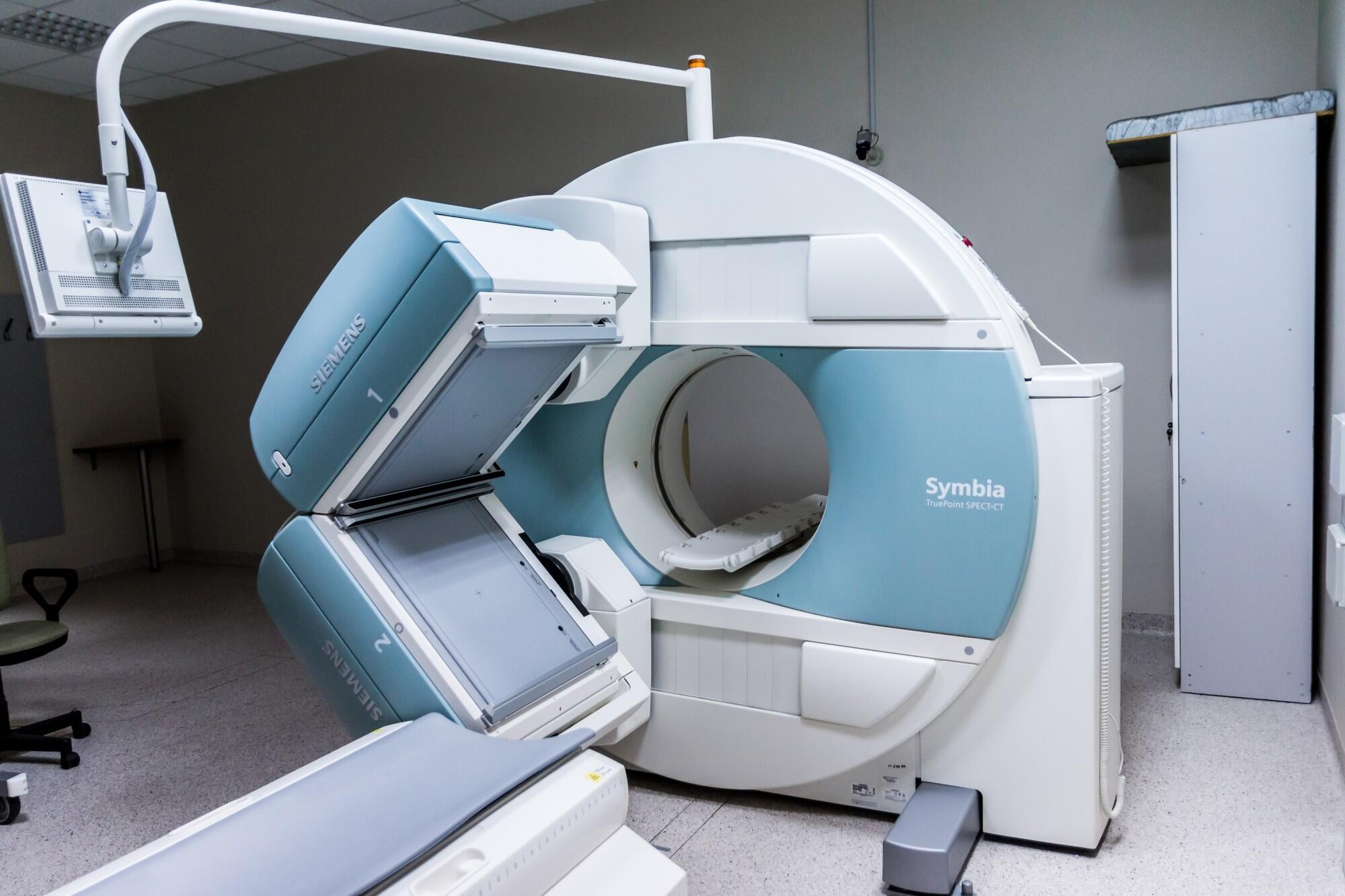
Musculoskeletal imaging is a crucial tool. It diagnoses and manages different conditions that affect the bones, muscles, joints, and soft tissues. With advancements in technology, there are now several types of musculoskeletal imaging techniques available.
In this guide, we will provide you with an informative and authoritative overview of the different types of musculoskeletal imaging and their functions.
X-Rays
X-rays are the most used method of musculoskeletal imaging. They use low levels of radiation to produce images of bones and other dense structures.
This includes calcium deposits or tumors. X-rays can help diagnose fractures, dislocations, arthritis, and bone infections.
You need X ray CE courses to be a licensed technician. This type of musculoskeletal imaging is quick, non-invasive, and cost-effective. But, it only provides 2-dimensional images and does not show soft tissues well.
Computed Tomography (CT)
A CT scan uses X-rays to create detailed cross-sectional images of the body. It is particularly useful for diagnosing complex fractures, joint problems, and soft tissue injuries.
CT scans are more detailed than X-rays. They can provide a 3-dimensional view of the musculoskeletal system.
But, they do expose patients to higher levels of radiation. They also may not be suitable for pregnant women or children.
Magnetic Resonance Imaging (MRI)
MRI uses powerful magnets and radio waves to create detailed images of bones, muscles, cartilage, and tendons. It is often used to diagnose sports injuries, spine problems, and joint disorders.
One of the main advantages of MRI is its ability to produce high-resolution images without using radiation. But, it can be expensive and time-consuming compared to other imaging techniques.
Ultrasound
Ultrasound uses sound waves to create real-time images of soft tissues. This includes muscles, ligaments, and tendons. It is particularly useful for diagnosing sprains, tears, and other types of soft tissue injuries.
Ultrasound is non-invasive and does not use radiation. This makes it a safe option for pregnant women and children.
But, it may not provide as detailed images as other techniques. It can also be operator-dependent.
Bone Scan
Bone scans involve injecting a small amount of radioactive material into the bloodstream. This substance then collects in areas where there is increased bone activity. This includes fractures, infections, or tumors.
Bone scans are sensitive and can detect abnormalities even before they are visible on X-rays. But, they do expose patients to a small amount of radiation.
Positron Emission Tomography (PET) Scan
A PET scan involves injecting a radioactive tracer into the body. This collects in areas with high metabolic activity. It is used to detect bone cancer or osteomyelitis (bone infection).
PET scans can provide detailed images of bones and soft tissues. This helps doctors determine if cancer has spread to other parts of the body. But, they also expose patients to higher levels of radiation.
Learn About the Types of Musculoskeletal Imaging
Musculoskeletal imaging plays a crucial role in the diagnosis and management of various conditions affecting bones, muscles, joints, and soft tissues. With advancements in technology, there are now several types of imaging techniques available, each with its benefits and limitations.
Healthcare professionals need to be informed about these options to choose the most appropriate one for each patient. So keep learning, stay informed, and take charge of your musculoskeletal health!
Is this article helpful? Keep reading our blog for more.


